

DIGITALIZATION OF DIAGNOSIS AND PREVENTION OF DUST-INDUCED LUNG DISEASES IN KEMEROVO COAL MINERS: INTEGRATION OF TELEMEDICINE AND BIG DATA ANALYTICS FOR PROTECTING THE HEALTH OF COAL INDUSTRY WORKERS
Main Article Content
Abstract
The implementation of digital technologies significantly improves the diagnosis, monitoring, and prevention of occupational diseases caused by chronic inhalation of coal dust in the coal industry. This paper presents the results of a study on cytological criteria that enable the identification of pulmonary dust lesions at various stages of their development. The use of modern digital platforms (telemedicine, big data, image analysis) and specialized software, such as the automated counting program «BioVision 4 series», is examined as contributing to more accurate and faster diagnostics, as well as improving the effectiveness of preventive measures. These results can be integrated into a digital medical examination support system, which significantly contributes to the modernization of occupational safety approaches and the innovative development of the coal industry.
Aim of the study is to evaluate the effectiveness of the «BioVision 4 series» program for diagnosing pneumoconiosis, as well as to conduct a comparative analysis of automated and traditional methods for diagnosing fibrotic changes and dust inclusions in lung tissue.
Materials and methods. The study involved cytological samples from 100 coal industry workers diagnosed with pneumoconiosis. The samples were processed using both traditional manual methods and the «BioVision 4 series» program. The main parameters for analysis were the area of fibrotic changes, the number of dust inclusions, and the level of inflammation in the lung tissue. All data were subjected to statistical processing to identify relationships between age, disease stage, and analysis results.
Results. The results of the study demonstrated that the automated method using the «BioVision 4 series» software significantly increases diagnostic accuracy, reducing both analysis time and the number of errors in counting cells and dust inclusions. It was also found that in the later stages of the disease, there is a marked increase in the area of fibrotic changes and the number of dust inclusions. The analysis of the obtained data indicates the high efficiency of the developed program in monitoring disease dynamics and facilitating early diagnosis. Additionally, the software enables the creation of a structured database with photo documentation, allows for the preservation and continuous updating of information during follow-up, and generates comparative reports. These features contribute to enhancing the informativeness of clinical assessments and support evidence-based diagnostic decision-making.
Conclusions. The use of the «BioVision 4 series» program for diagnosing pneumoconiosis is a highly effective tool that provides more accurate and faster results compared to traditional methods. This opens new possibilities for early diagnosis, monitoring the health status of coal industry workers, and improving medical services in this field.
Keywords
Author Biographies
Олег Иванович Бондарев,
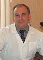
Oleg I. Bondarev
MD, PhD, head of the research laboratory for pathological anatomy
Сергей Николаевич Филимонов,
доктор мед. наук, профессор, начальник отдела экологии человека, общественного здоровья и здравоохраненияArticle Details
Information about financing and conflict of interests
The authors declare that they have no apparent or potential conflicts of interest related to the publication of this article.
 This work is licensed under a Creative Commons Attribution 4.0 License.
How to Cite
References
Butenko GR, Plyakin VA, Polonskaya NYu, Volchenko NN, Glukhova YuK, Trubitsyn MA. Development of clinical cytology as a medical specialty in Russia. P.A. Herzen Journal of Oncology. 2023; 12(5): 39-44. Russian (Бутенко Г.Р., Плякин В.А., Полонская Н.Ю., Волченко Н.Н., Глухова Ю.К., Трубицын М.А. Развитие клинической цитологии как медицинской специальности в России //Онкология. Журнал имени П.А. Герцена. 2023. Т. 12, № 5. С. 39-44.) doi: 10.17116/onkolog20231205139
Lelevich SV, Anisovich EI. Cytological Studies in Medicine. Medical News. 2018; 2: 33-35. Russian (Лелевич С.В., Анисович Е.И. Цитологические исследования в медицине //Медицинские новости. 2018. № 2. С. 33-35)
Tkachuk E.A., Seminsky I.Zh. The Role of Genetics in Modern Medicine. Baikal Medical Journal. 2022; 1(1): 81-88. Russian (Ткачук Е.А., Семинский И.Ж. Роль генетики в современной медицине //Байкальский медицинский журнал. 2022.
Т. 1. № 1. С. 81-88. doi: 10.57256/2949-0715-2022-1-81-88
Panov YuP, Grabskiy AA, Rozhkov AA. Current Status and Prospects for the Development of Digital Technologies in the Russian Coal Industry. Proceedings of Higher Educational Establishments: Geology and Exploration. 2023; (5): 8-21. Russian (Панов Ю.П., Грабский А.А., Рожков А.А. Современное состояние и перспективы развития цифровых технологий в угольной промышленности России //Известия высших учебных заведений. геология и разведка. 2023. № 5. С. 8-21.) doi: 10.32454/0016-7762-2023-65-5-8-21
Solovenko IS, Rozhkov AA, Lizunkov VG, Malushko EYu. Digitalization of Coal Industry Enterprises in Russia: Problem Statement. Questions of History. 2022; 5(2): 152-165. Russian (Соловенко И.С., Рожков А.А., Лизунков В.Г., Малушко Е.Ю. Цифровизация предприятий угольной промышленности России: к постановке проблемы //Вопросы истории. 2022. № 5(2). С. 152-165)
Lebedev GS, Shaderkin IA, Tertychny AS, Shaderkina AI. Digital Pathomorphology: Creating an Automated Microscopy System. Russian Journal of Telemedicine and e-Health. 2021; 7(4): 27-47. Russian (Лебедев Г.С., Шадеркин И.А., Тертычный А.С., Шадеркина А.И. Цифровая патоморфология: создание системы автоматизированной
микроскопии //Российский журнал телемедицины и электронного здравоохранения. 2021;7(4);27-47.) doi: 10.29188/2712-9217-2021-7-4-27-47
Montironi R, Cimadamore A, Scarpelli M, Cheng L, Lopez-Beltran A, Mikuz G. Pathology Without a Microscope: From Projection Screen to Virtual Slide. Pathology – Research and Practice. 2020; 216(11): 153196. Russian (Монтирони Р., Чимадаморе А., Скарпелли М., Чэн Л., Лопес-Бельтран А., Микуз Д. //Патология без микроскопа: от проекционного экрана до виртуального слайда //Pathology – Research and Practice. 2020. Т. 216, № 11. С. 153196.) doi: 10.1016/j.prp.2020.153196
Borowsky AD, Glassy EF, Wallace WD, Kallichanda NS, Behling CA, Miller DV, et al. Digital Whole Slide Imaging Compared With Light Microscopy for Primary Diagnosis in Surgical Pathology. Arch Pathol Lab Med. 2020; 144(10): 1245-1253. doi: 10.5858/arpa.2019-0569-OA
Fayyaz SM, Yaqub U, Hassan A, Munir MK, Iqbal S, Hamza M, Tahir FN. Role of AI and Machine Learning in Diagnosis and Management of Pulmonary, Gynecological and Dermatological Conditions. African Journal of Biological Sciences. 2024; 6(15): 12800-12811. doi: 10.48047/AFJBS.6.15.2024.12800-12811
World Health Organization (WHO). (2020). Telemedicine in remote areas: Strengthening health systems through digital innovation. World Health Organization. https://www.who.int/news-room/fact-sheets/detail/telemedicine